icobrain ms
Accurately visualize lesion and brain volume changes over time in multiple sclerosis
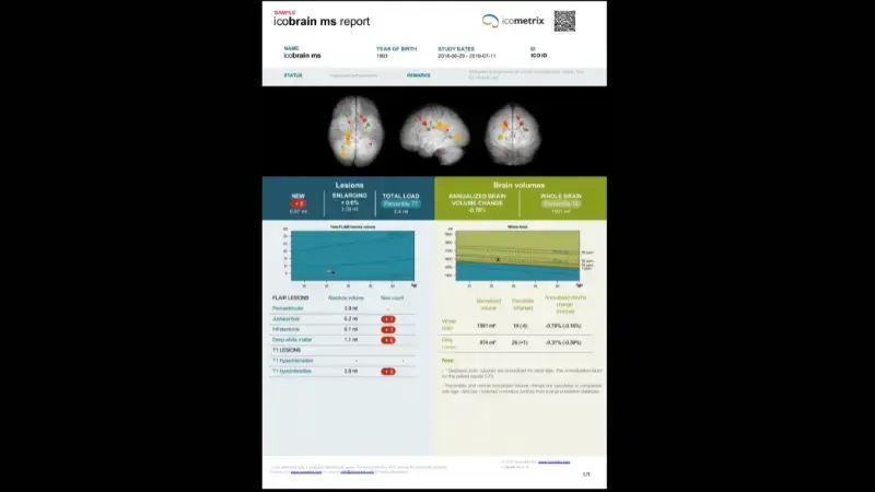
icobrain ms is designed to support the detection and tracking of disease progression, and the evaluation of treatment effectiveness. It provides a quantitative analysis of the inflammatory component of the disease, assessing longitudinal changes in FLAIR and T1 lesions. Additionally, icobrain ms can be used to assess changes in brain volume over time, which can be compared to age- and sex-matched healthy individuals.

Assesses lesion dissemination in space and time.
Detecting and quantifying FLAIR white matter hyperintensities, T1 hypointensities, and contrast-enhancing lesions to evaluate disease activity. Tracking changes in FLAIR lesion number and volume to monitor progression. Comparing FLAIR lesion volume to a reference population
Provides brain volume change metrics
Tracking annualized changes in whole brain and gray matter volume. Comparing brain volumes to age- and sex-matched normative data and a clinical practice reference population
Detect disease progression
Improved detection
Adopt clinical practice guidelines
Save time, secure your diagnosis and optimize your workflow with Incepto
Publications
-
Jain S., Ribbens A., Sima D., Cambron M., De Keyser J., Wang C., Barnett M., Van Huffel S., Maes F., Smeets D., Two Time Point MS Lesion Segmentation in Brain MRI: An Expectation Maximization Framework, Frontiers in Neuroscience: Brain Imaging Methods, 2016 Dec 19;10:576 (https://www.frontiersin.org/articles/10.3389/fnins.2016.00576/full)
-
Mettenburg JM, Branstetter BF, Wiley CA, Lee P, Richardson RM. Improved Detection of Subtle Mesial Temporal Sclerosis: Validation of a Commercially Available Software for Automated Segmentation of Hippocampal Volume. AJNR Am J Neuroradiol. 2019 Mar;40(3):440-445. doi: 10.3174/ajnr.A5966.
-
Akaishi T, Nakashima I, Mugikura S, Aoki M, Fujihara K. Whole brain and grey matter volume of Japanese patients with multiple sclerosis. J Neuroimmunol. 2017 May 15;306:68-75. doi: 10.1016/j.jneuroim.2017.03.009. Epub 2017 Mar 16. PMID: 28385190.
-
Akaishi T, Takahashi T, Fujihara K, Misu T, Mugikura S, Abe M, Ishii T, Aoki M, Nakashima I. Number of MRI T1-hypointensity corrected by T2/FLAIR lesion volume indicates clinical severity in patients with multiple sclerosis. PLoS One. 2020 Apr 3;15(4):e0231225. doi: 10.1371/journal.pone.0231225. PMID: 32243459; PMCID: PMC7122737.
-
Beadnall HN, Wang C, Van Hecke W, Ribbens A, Billiet T, Barnett MH. Comparing longitudinal brain atrophy measurement techniques in a real-world multiple sclerosis clinical practice cohort: towards clinical integration? Ther Adv Neurol Disord. 2019 Jan 25;12:1756286418823462. doi: 10.1177/1756286418823462. PMID: 30719080; PMCID: PMC6348578.
Regulatory
Contact us to know if this product is available in your country.

