icobrain tbi
Quantify traumatic brain injury and track structural changes
icobrain tbi for CT can be used as part of the assessments of the acute impacts of brain trauma, reporting the mass effect - the volumes of hyperdensities, cisterns, and ventricles, as well as the midline shift. icobrain tbi for MRI can be used as part of the assessments of sub-acute and longitudinal impacts of brain trauma, especially traumatic axonal injuries, by reporting on subtle anomalies in brain volume and inflammation through longitudinal monitoring.

For CT uncovers mass effects
Management of traumatic axial injuries and atrophy in patients with mild to severe brain trauma. Detect, quantify, and classify hyperdensities; epidural; subdural and intra-parenchymal.
Assess the ventricular and cisternal CSF spaces and ventricular asymmetry. Measure midline shift. Compare volumes to an age-and sex matched normative reference population.
For MRI uncovers diffuse consequences of brain damage
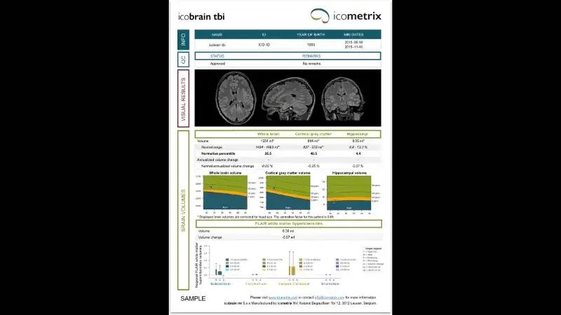
Fast, standardized and objective management of acute brain trauma. Detect, quantify, and track traumatic axonal injuries (TAI)
Assess TAI distribution by Adams-Gentry Classification (subcortical, cerebellum, corpus callosum, brainstem). Track annual changes in cortical and hippocampal volumes. Compare brain volumes to an age- and sex-matched normative population.
Automate the identification, labeling, and quantification of segmentable brain structures
Reduce subjectivity of human observers
Save time, secure your diagnosis and optimize your workflow with Incepto
Publications
-
Ferrazzano P, Yeske B, Mumford J, Kirk G, Bigler ED, Bowen K, O'Brien N, Rosario B, Beers SR, Rathouz P, Bell MJ, Alexander AL. Brain Magnetic Resonance Imaging Volumetric Measures of Functional Outcome after Severe Traumatic Brain Injury in Adolescents. J Neurotrauma. 2021 Jun 1;38(13):1799-1808. doi: 10.1089/neu.2019.6918. Epub 2021 Feb 24. PMID: 33487126; PMCID: PMC8219192.
-
Bohyn C, Vyvere TV, Keyzer F, Sima DM, Demaerel P. Morphometric evaluation of traumatic axonal injury and the correlation with post-traumatic cerebral atrophy and functional outcome. Neuroradiol J. 2022 Aug;35(4):468-476. doi: 10.1177/19714009211049714. Epub 2021 Oct 13. PMID: 34643120; PMCID: PMC9437508.
-
Jain S, Vyvere TV, Terzopoulos V, Sima DM, Roura E, Maas A, Wilms G, Verheyden J. Automatic Quantification of Computed Tomography Features in Acute Traumatic Brain Injury. J Neurotrauma. 2019 Jun;36(11):1794-1803. doi: 10.1089/neu.2018.6183. Epub 2019 Feb 1. PMID: 30648469; PMCID: PMC6551991.
-
Vande Vyvere T, Pisică D, Wilms G, Claes L, Van Dyck P, Snoeckx A, van den Hauwe L, Pullens P, Verheyden J, Wintermark M, Dekeyzer S, Mac Donald CL, Maas AIR, Parizel PM. Imaging Findings in Acute Traumatic Brain Injury: a National Institute of Neurological Disorders and Stroke Common Data Element-Based Pictorial Review and Analysis of Over 4000 Admission Brain Computed Tomography Scans from the Collaborative European NeuroTrauma Effectiveness Research in Traumatic Brain Injury (CENTER-TBI) Study. J Neurotrauma. 2024 Oct;41(19-20):2248-2297. doi: 10.1089/neu.2023.0553. Epub 2024 Apr 18. PMID: 38482818.
Regulatory
Contact us to know if this product is available in your country.

